


您的位置:首页 > 产品中心 > Anti-Peripherin-2 Antibody, clone 5H2
Anti-Peripherin-2 Antibody, clone 5H2
产品别名
Anti-Peripherin-2 Antibody, clone 5H2
Retinal degeneration slow protein
基本信息
| eCl@ss | 32160702 |
| NACRES | NA.41 |
| General description【一般描述】 | Peripherin-2 (UniProt: P17810; also known as Retinal degeneration slow protein) is encoded by the PRPH2 (also known as RDS) gene in bovine species. Peripherin-2 is a disulfide-linked homodimeric, multi-pass membrane protein that may function as an adhesion molecule involved in stabilization and compaction of outer segment disks in retina and is considered to be essential for disk morphogenesis. It is exclusively expressed in outer segments. Peripherin-2 can form homo- and heteromeric protein complexes in outer segment. The core homomeric peripherin-2 unit is the non-covalent tetramer, which can also assemble into covalently linked octamers and higher-order oligomers. The homomeric peripherin-2 complexes are essential for proper outer segment morphology and architecture. Peripherin-2 is also shown to form heteromeric complexes with its homolog ROM-1. Mutations in PRPH2 gene can lead to autosomal dominant retinitis pigmentosa (adRP) And a majority of these mutations are located within the large loop domain connecting the transmembrane domain 3 and 4 (Ref.: Bohm, S et al. (2017). Scientific Reports 7, Article number: 2321). |
| Specificity【特异性】 | Clone 5H2 detects Peripherin-2 in bovine and mouse retina. |
| Immunogen【免疫原】 | Purified bovine whole peripherin. Epitope: C-terminus |
| Application【应用】 | Anti-Peripherin-2, clone 5H2, Cat. No. MABN2435, is a highly specific mouse monoclonal antibody that targets Peripherin-2 and has been tested in Immunofluorescence, Radioimmunoassay, and Western Blotting. Research Category Neuroscience Western Blotting Analysis: A representative lot detected Peripherin-2 in Western Blotting applications (Connell, G., et. al. (1991). Proc Natl Acad Sci USA. 88(3):723-6; Zhang, Y., et. al. (2009). J Cell Sci. 122(Pt 8):1192-200). Radioimmunoassay Analysis: A representative lot detected Peripherin-2 in Radioimmunoassay applications (Connell, G., et. al. (1991). Proc Natl Acad Sci USA. 88(3):723-6). Immunofluorescence Analysis: A representative lot detected Peripherin-2 in Immunoflourescence applications (Zhang, Y., et. al. (2009). J Cell Sci. 122(Pt 8):1192-200). |
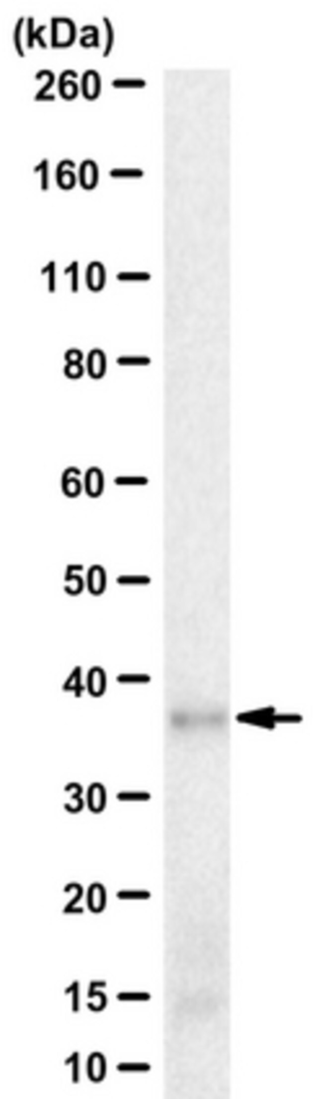
| Quality【质量】 | Evaluated by Western Blotting in bovine retinal tissue lysate. Western Blotting Analysis: 4 µg/mL of this antibody detected Peripherin-2 in 10 µg of bovine retinal tissue lysate. |
| Physical form【外形】 | Protein G purified Purified mouse monoclonal antibody IgG1 in buffer containing 0.1 M Tris-Glycine (pH 7.4), 150 mM NaCl with 0.05% sodium azide. Format: Purified |
| Other Notes【其他说明】 | Concentration: Please refer to lot specific datasheet. |
产品性质
| Quality Level【质量水平】 | 100 |
| biological source【生物来源】 | mouse |
| antibody form【抗体形式】 | purified immunoglobulin |
| antibody product type | primary antibodies |
| clone【克隆】 | 5H2, monoclonal |
| species reactivity | bovine, mouse |
| packaging【包装】 | antibody small pack of 25 μg |
| technique(s) | immunofluorescence: suitable radioimmunoassay: suitable western blot: suitable |
| isotype【同位素/亚型】 | IgG1κ |
| UniProt accession no.【UniProt登记号】 | P17810 |
| shipped in【运输】 | ambient |
产品说明
| Target description【目标描述】 | ~39 kDa observed; 38.99 kDa calculated. Uncharacterized bands may be observed in some lysate(s). |
| Storage and Stability【储存及稳定性】 | Stable for 1 year at 2-8°C from date of receipt. |
| Disclaimer【免责声明】 | Unless otherwise stated in our catalog or other company documentation accompanying the product(s), our products are intended for research use only and are not to be used for any other purpose, which includes but is not limited to, unauthorized commercial uses, in vitro diagnostic uses, ex vivo or in vivo therapeutic uses or any type of consumption or application to humans or animals. |